Shoulder – Proximal Humerus Fracture
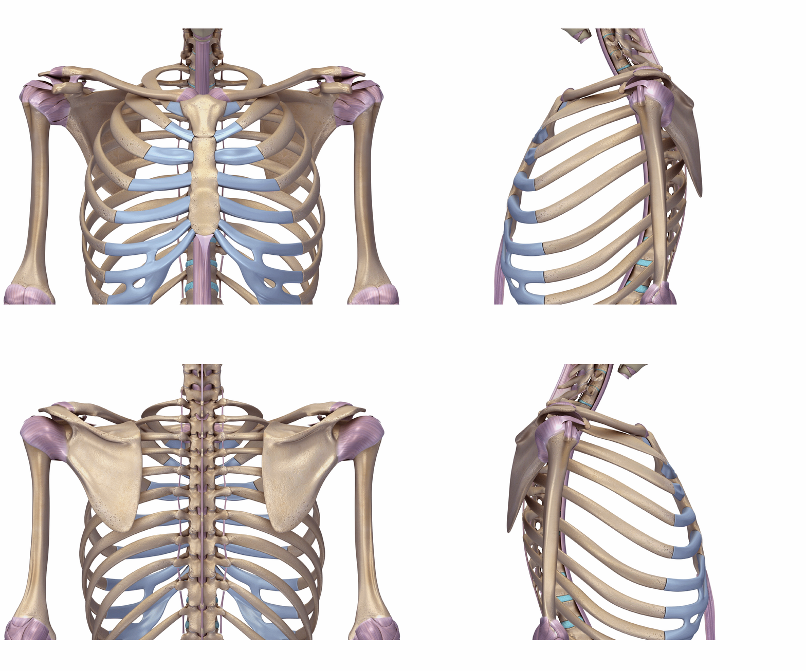
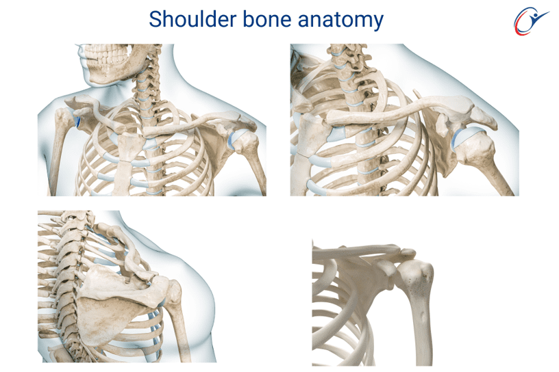
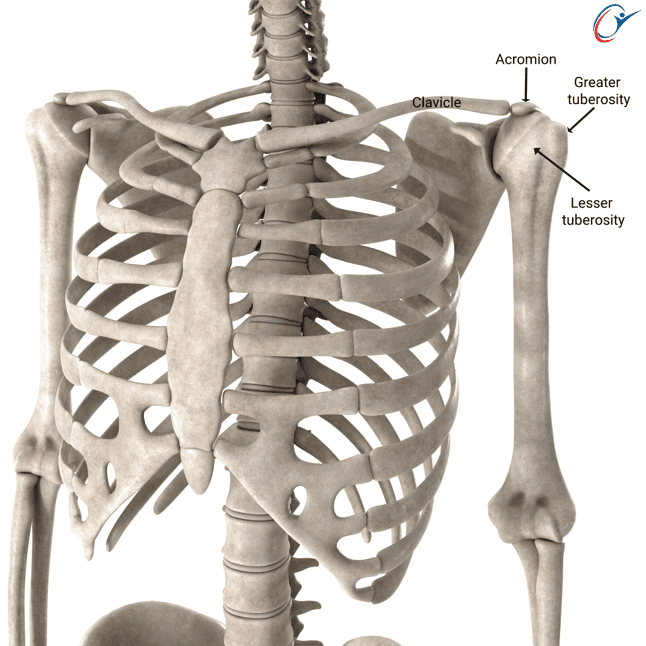
Shoulder anatomy
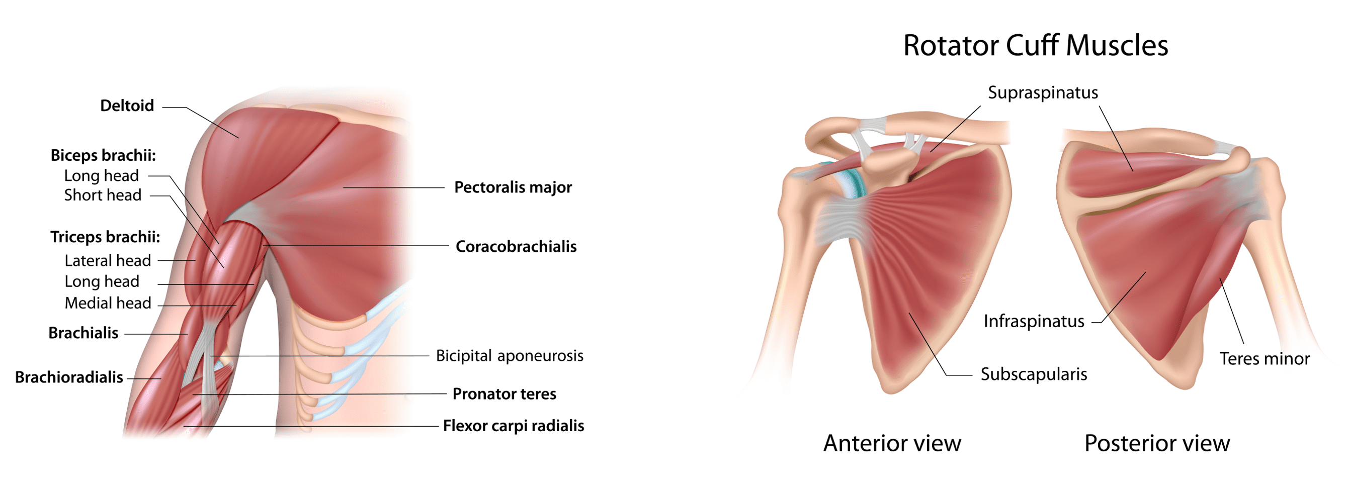
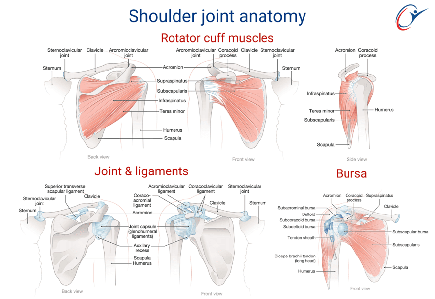
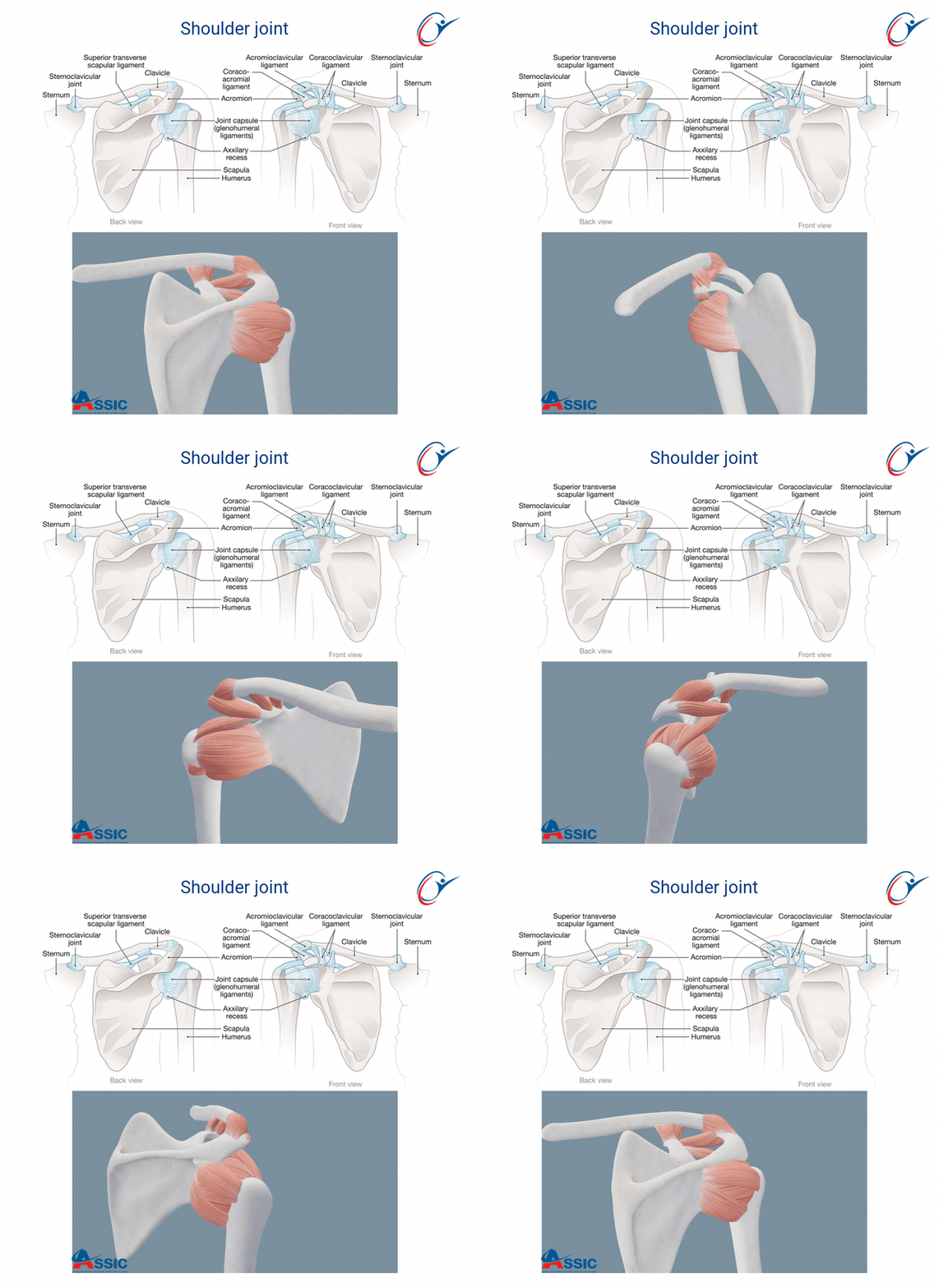
- The shoulder joint is a multi-axial joint with muscles crossing it to allow shoulder range of motion (ROM)
- The rotator cuff muscles are important in allowing shoulder ROM
- The rotator cuff muscles consists of Subscapularis, Supraspinatus, Infraspinatus and Teres Minor
- Other important shoulder muscles that allow shoulder ROM include long head of Biceps and the Deltoid muscle


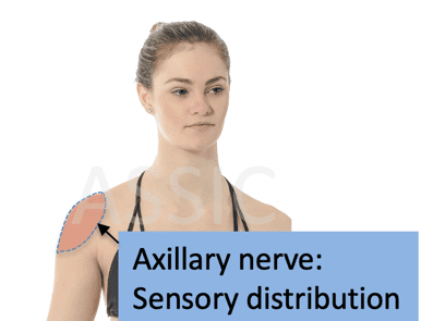
Axillary nerve function
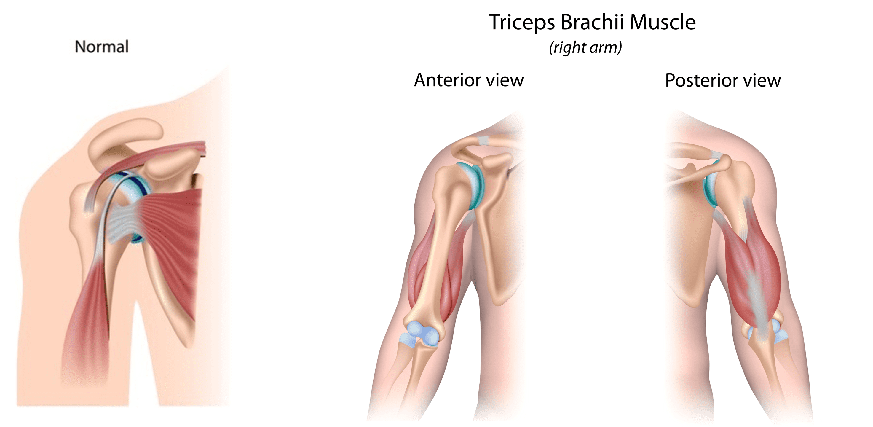
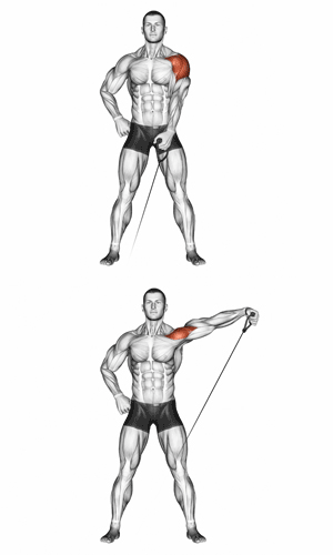
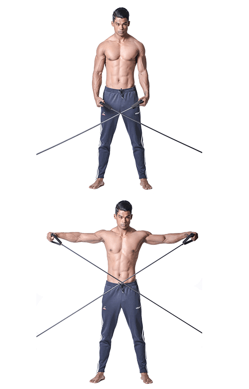
The Axillary nerve supplies the Deltoid muscle & the muscle is responsible for shoulder abduction movement. In clinical practice to test nerve motor function: test resisted shoulder abduction with arm at 90° abducted position.

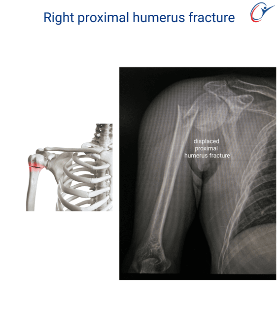
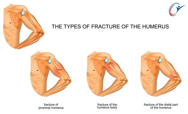
Proximal humerus fracture
The mechanism usually a fall to the arm or motor vehicle accident
More common in females
Risk factors:
Osteoporosis
Presentation:
- Pain
- Swelling
- Deformity
- Decreased Range Of Motion (ROM)
Clinically:
- Swelling and bruising of chest, arm, forearm
- Bone tenderness
- Assess axillary nerve and arterial function and exclude associated injury
Investigations:
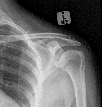
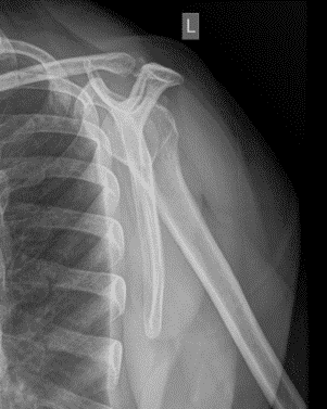
- X-Ray: AP, axillary and lateral
- CT: for fracture evaluation and surgical planning
Anterior-posterior view shoulder view
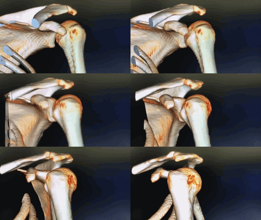
Lateral shoulder view
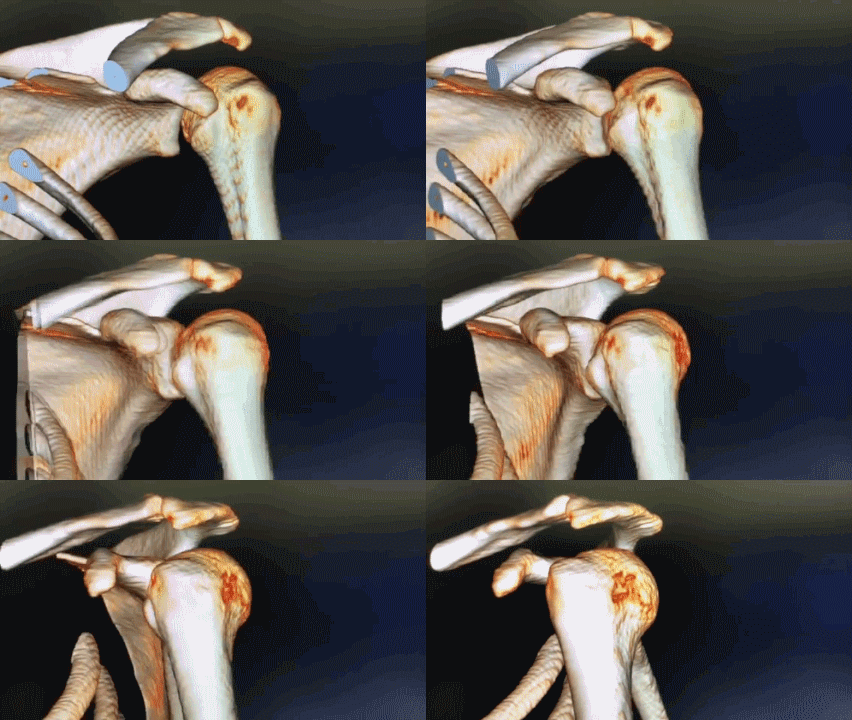
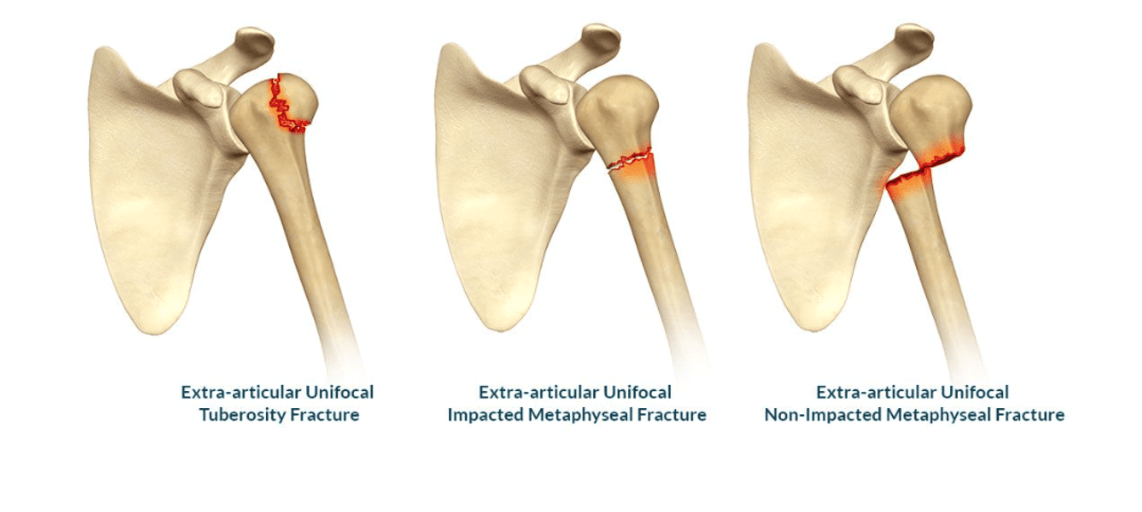
Extra-articular shoulder fractures


Neers classification for proximal humerus fractures
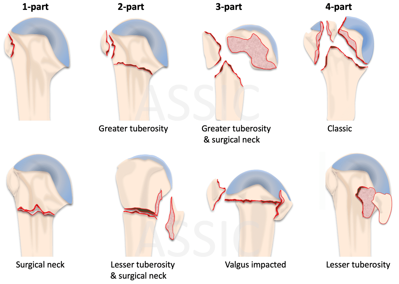
AO classification of proximal humerus fractures

Hertel criteria for predicting avascular necrosis of humeral head fracture
Proximal humerus fracture risk factors for AVN include:
- Medial calcar segment < 8mm
- Medial hinge disruption (>2 mm displacement of the diaphysis)
- Medial metaphysis comminution
- Anatomic neck fracture
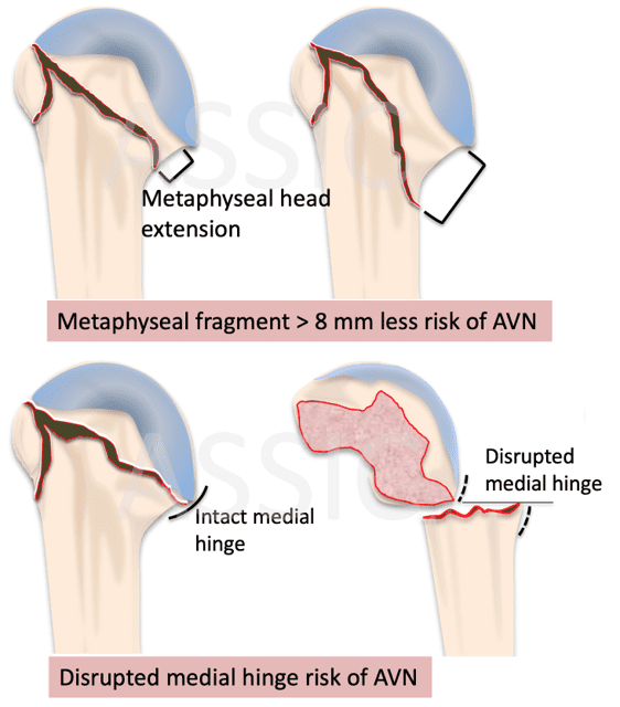
Tuberosity fracture management
Nondisplaced greater tuberosity fracture can be managed nonoperatively with immobilization.
Arm sling and wrath/collar and cuff/shoulder immobilizer for 2-3 weeks
- Active forearm and Hand grip exercises
- Pendulum shoulder exercise as pain permits
- Scapular stabilizer exercises


Displaced greater tuberosity fractures
Surgery indications:
- > 1cm displacement
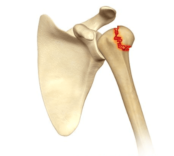
Greater Tuberosity Fracture
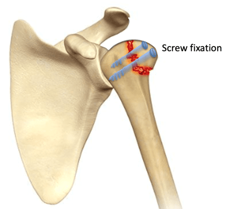
Greater Tuberosity Surgical Screw Fixation
Proximal Humerus head/neck fractures
Surgery indications:
- Displaced
Options:
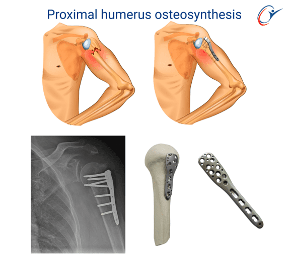
- ORIF plate & screw fixation
- K-wire fixation
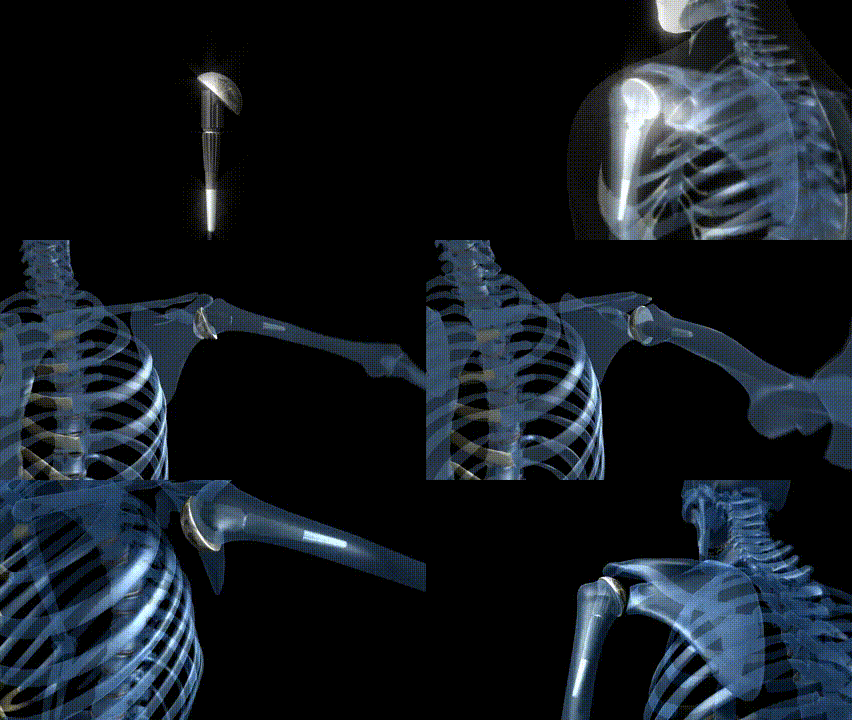
- Shoulder joint replacement
Proximal humerus fracture plate and screw fixation
Shoulder joint replacement principles

Surgery Technique



Surgery Greater tuberosity screw fixation
Approach: Deltoid
Fixation: screw fixation

Proximal humerus fracture plate and screw fixation
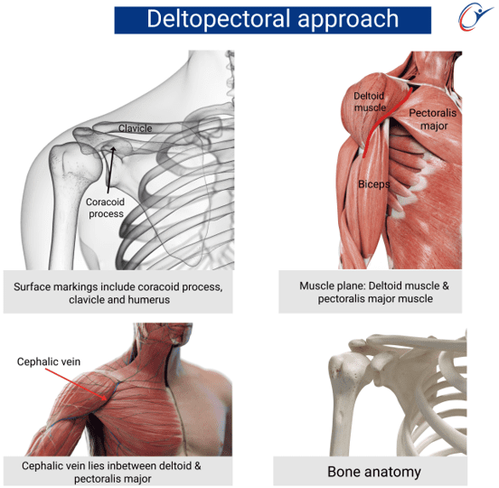
Deltopectoral approach
Plate and screw fixation
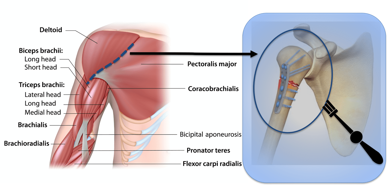
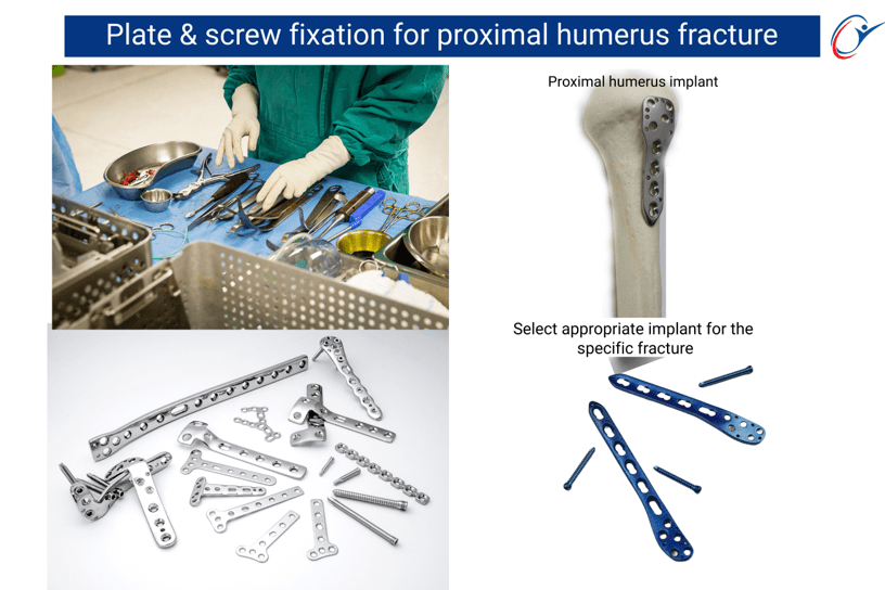

Shoulder joint replacement
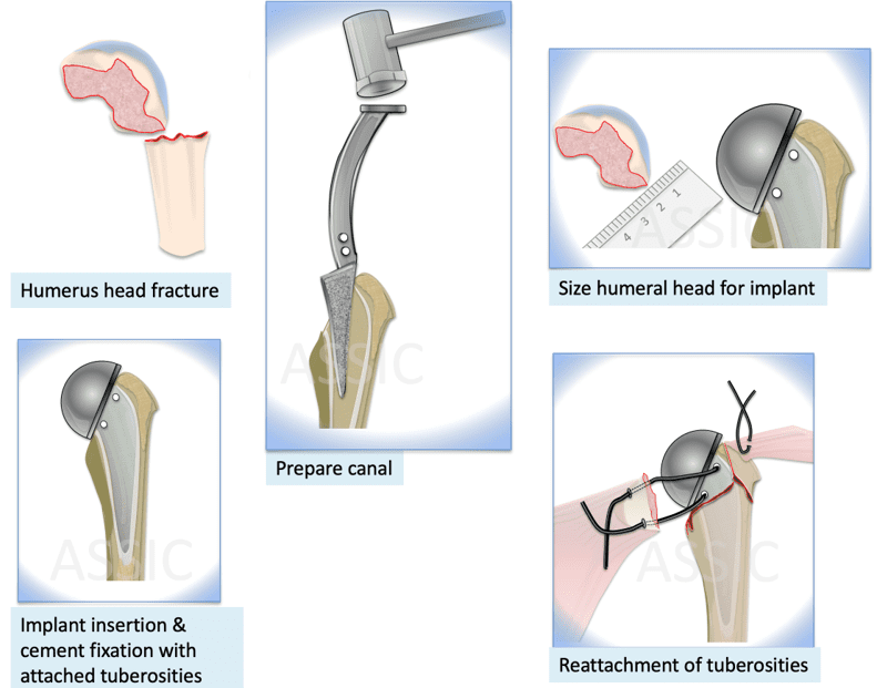
Principles of shoulder joint replacement:
Approach: Deltopectoral
Arthroplasty:
- Bone template cuts
- Prepare humerus shaft
- Size humerus head for implant size selection
- Suture tuberosities for fixation to implant
- Implant insertion
Rehabilitation
Phases of rehabilitation:
- Immobilization
- Passive/ assisted ROM/isometric exercise of shoulder girdle
- Progressive strengthening exercises with resistance exercises after 6-8 weeks
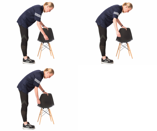
Pendulum Shoulder Exercise

Hand Grip Exercise


References
- Fractures of the proximal humerus in osteoporotic bone. Ralph Hertel. Osteoporosis Int (2005) 16: S65-S72.
- Complex proximal humerus fractures: Hertel’s criteria reliability to predict head necrosis. G Campochiaro, M Rebuzzi, P Baudi, F Catani. Musculoskelet Surg. 2015 Sep;99 Supl 1: S9-15.
Contributors
Dr J Kauta
Dr M Y Hassan
Prof S Maqungo