Ankle & Foot – Osteochondral Talus Lesions
Lower limb/ankle anatomy
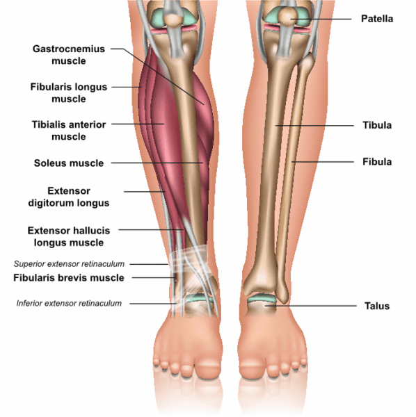
The talocrural (ankle) joint is the junction of three bones: distal ends of tibia and fibular and the talus trochlear
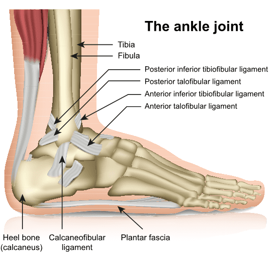
The tibia and fibular are bound by the ligamentous structures that include the syndesmosis, anterior/posterior/transverse tibiofibular ligaments.
Collateral ligaments include the anterior and posterior fibulotalar ligaments, fibulocalcanear on the lateral side while the medial side is supported by the deltoid ligament and calcaneonavicular ligaments.
The ankle joint is not a pure hinge joint as there is some rotation around the helical axis of the joint due to the asymmetrical shape of the talus


Precise ankle joint congruence is important for load distribution of the compressive forces across the joint
Ankle sprain/fractures and osteochondral lesions
The ankle joint consists of the distal end of the fibular and tibia. The fibular and posterior/medial malleoli of the tibia articulates with the talus bone thereby completing the ankle joint with supporting ligaments. Injury and fractures of the ankle joint implies breakage of these related structures.
Ankle ligament/s and/or fractures commonly occur.
Occasional osteochondral talus fractures are associated with ankle sprains/fractures.
Causes:
- Twisting/rotational movements with ankle inversion mechanism during activity, especially during sport activities
Presentation:
- Pain
- Swelling
- Mechanical problems like clicking/locking
- Chronic cases may present with intermittent ankle swelling ± associated ankle mechanical catching symptoms
Clinically:
- Assess ankle ligament stability
- Bone tenderness over the lesions with posteromedial tenderness behind the medial malleolus with ankle in dorsiflexion while anterolateral lesions having anterolateral bone tenderness when ankle in maximum plantarflexion
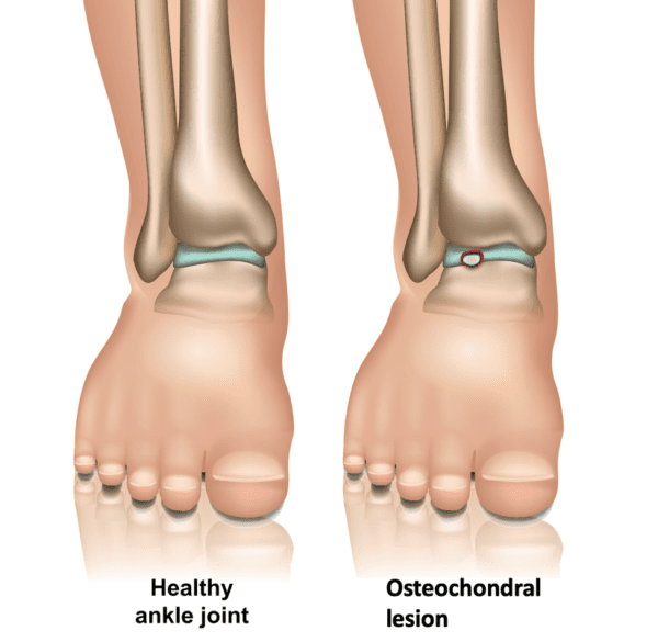
Talus osteochondral lesion (OC)
OC lesions may be on medial or lateral side
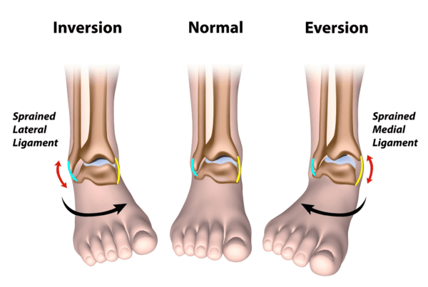
Ankle injury mechanism
Injury Mechanism:
- Ankle inversion or eversion movement ±rotational component. Either the ligament is torn or the ankle is fractured
- Ankle inversion injury associated with Talus osteochondral lesions
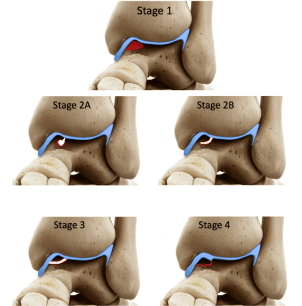
Ferkel CT classification of talus OC lesions
Ⅰ: Cystic lesion with roof intact
ⅡA: Cystic lesion communicating with talar dome
ⅡB: Open articular surface lesion overlying the undisplaced fragment
Ⅲ: Undisplaced lesion with lucency
Ⅳ: Displaced fragment
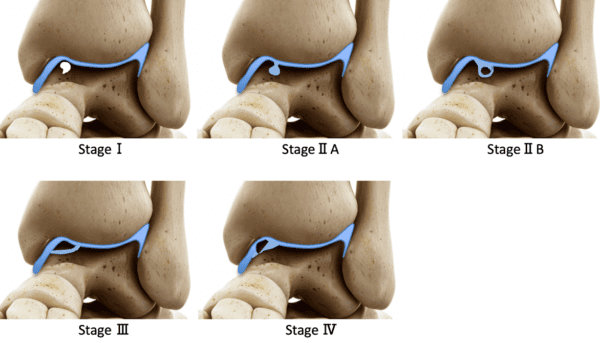
Berndt & Harty classification of talus OC lesions
Stage Ⅰ: Subchondral bone compression with marrow oedema
Stage Ⅱ
- Stage Ⅱa: Subchondral cyst
- Stage Ⅱb: Incomplete fragment separation
Stage Ⅲ: complete fragment separation but no displacement
Stage IV : Displaced fragment
Investigations
X-Rays may show the talus osteochondral lesion
CT scan: useful for assessment of the bony component of the OC lesion and for cystic change evaluation. Useful for preoperative planning
MRI scan useful to identify osteochondral lesion and assess lesion stability together with assessing any associated bone bruising and/or ligamentous injury
Treatment principles
Non-operative:
- immobilization with brace/cast with non-weight bearing for associated ankle fractures and stable OC lesions (acute, nondisplaced) that require conservative treatment
Surgery:
- Displaced osteochondral lesions
- Large lesions
Surgery options:
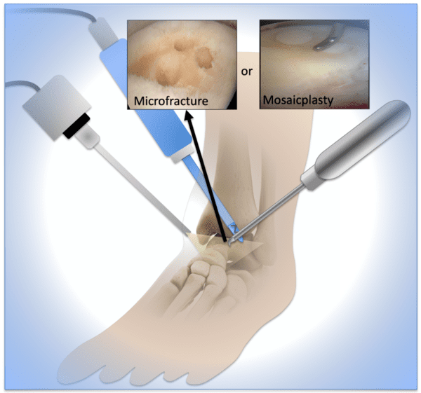
Ankle arthroscopic removal of loose body ± microfracture for lesions < 1cm
OC grafting
- Mosaicplasty
- Allograft implantation
- Autologous chondrocyte implantation
Surgery Technique

Ankle arthroscopy

Ankle arthroscopy procedure
Removal of OC lesions
± microfracture
± mosaicplasty
± autologous chondrocyte implantation
Surgery: Talus microfracture or mosaicplasty
Match readiness
Return to sport & match readiness:
- After appropriate rehabilitation
- Assess return to sport after completing battery of testing

Foot Strengthening Exercise

Resistance Strength Exercise

Ankle Strapping
Download ASSIC performance fingerprint app or ASSIC strength/conditioning app for ankle rehab guidelines and strapping technique under professional supervision
References
- Fractures of the ankle joint. Hans Goost, Matthias D Wimmer, Alexej Barg, Kouroush Kabir, Victor Valderrabano, Christof Burger. Dtsch Arztebl Int. 2014 May;111(21): 377-388.
- Osteochondral lesions of the talus. RD Santrock, MM Buchanan, TH Lee, GC Berlet. Foot Ankle Clin N Am;8(2003): 73-90.
- Osteochondral lesions of the talus: Current concept. O Laffenêtre. Orthop Traumatol Surg res. 2010 Sep;96(5): 554-66.
- Osteochondral lesions of the talus: a new magnetic resonance grading system with arthroscopic correlation. DN Mintz, GS Tashjian, DA Connell, JT Deland, M O’Malley, HG Potter. Arthroscopy. 2003 Apr;19(4): 353-9.
Contributors
Dr M Y Hassan
Dr C Marais